Dental X-Rays

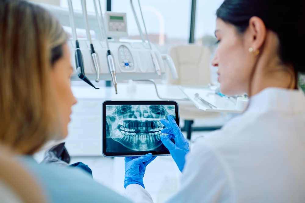
Hidden problems inside your teeth and jaw rarely announce themselves early. That’s why Dr. Adam Foleck, DMD, and the team at The Foleck Center rely on dental X-rays, also called radiographs, to look beneath the surface of your smile before small issues turn into big ones. Imaging tests let your dentist see what’s happening inside your teeth, gums, and jawbone. These are details a clinical exam alone can’t reveal, including decay, infection, bone loss, cysts, and changes in bone density in their earliest stages.

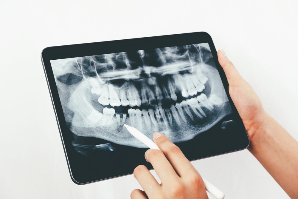
Different types of X-rays serve different purposes. Bitewing X-rays check for cavities and monitor filling fit, periapical X-rays capture an entire tooth from crown to root, and panoramic X-rays provide a full view of your upper and lower jaw in a single image. Occlusal X-rays focus on jaw alignment and developing teeth, while cephalometric and extraoral X-rays evaluate how the teeth and jaw relate to one another. Your dentist chooses the right type based on your specific oral health needs and history, so no two patients receive exactly the same imaging plan.

Does something feel off, but you can’t tell why?

Pain and sensitivity rarely point to just one cause, and guessing at the source can mean treating the wrong problem. The questions below help pinpoint whether imaging is what you actually need next.

Is a dental x-ray right for you?

Tooth pain or sensitivity can come from a dozen different sources, and many of them stay hidden beneath the surface where a visual exam alone can’t reach. Before deciding on treatment, it helps to know whether imaging is the right next step for finding what’s actually going on.

Benefits of Dental X-Rays

Imaging does more than confirm a suspicion, it often reveals problems long before you’d feel them. Here’s what that added visibility means for your care.

Early Detection of Hidden Decay

Cavities often form between teeth or beneath old fillings where they’re invisible to the naked eye. Dental X-rays catch this decay in its earliest stages, before it spreads deeper into the tooth or causes pain. Catching cavities early means simpler, less invasive treatment, like a standard filling instead of a root canal. It also helps you avoid the discomfort and expense of a more advanced dental problem down the road.

Accurate Treatment Planning

Clear, detailed images give your dentist the information needed to design a treatment plan that fits your exact situation. X-rays show root position, bone structure, and the relationship between teeth, which helps guide decisions about implants, braces, or extractions. This level of detail reduces guesswork and supports more predictable, successful outcomes for whatever care you need.

Monitoring Changes Over Time

Your oral health shifts gradually, and some changes are only visible when compared to earlier images taken over time. Regular X-rays let your dentist track bone density, monitor root development, and follow the progress of previous dental work year after year. This ongoing comparison makes it easier to spot subtle shifts before they turn into noticeable problems that require more involved treatment.

Lower Radiation with Digital Imaging

Modern digital X-rays use significantly less radiation than traditional film, making the process safer and more efficient. Images appear on screen within seconds, so there’s no waiting around for film to develop. Lead aprons and thyroid collars add another layer of protection during every scan. This combination keeps exposure to a minimum while still delivering sharp, reliable results.

Better Diagnosis of Gum and Bone Health

Gum disease often damages the bone supporting your teeth long before symptoms become obvious during a routine exam. X-rays reveal this bone loss clearly, allowing your dentist to catch periodontal issues early and recommend treatment before teeth become loose or unstable. This added insight protects both the appearance of your smile and your long-term oral health for years ahead.

What to Expect During Your Dental X-Ray Visit

Every visit follows a clear, patient-centered process from start to finish. Here’s what each step looks like, from your first conversation to your final treatment plan.

Talking Through Your Symptoms and History

Your visit starts with a short conversation about any pain, sensitivity, or concerns you’ve noticed. Dr. Adam Foleck asks about your dental history and reviews when you last had imaging done. This context shapes every decision that follows, so nothing about your visit feels routine or rushed.

Choosing the Right Type of X-Ray for You

Not every concern calls for the same image. Dr. Foleck selects between bitewing, periapical, panoramic, or other X-rays based on what he needs to see and where. A patient checking for cavities won’t need the same imaging as one being evaluated for an implant or a jaw concern.

Capturing Clear Images in Minutes

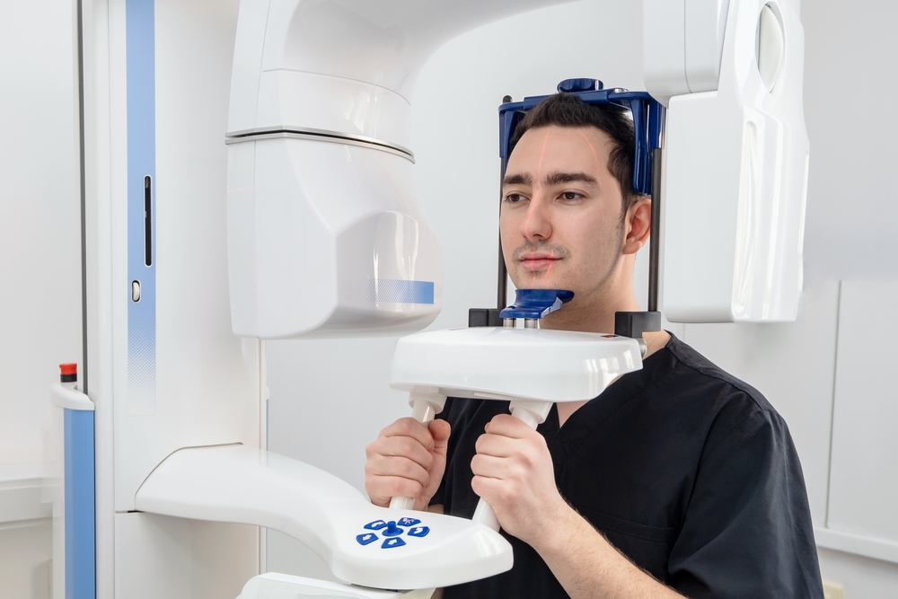
A technician positions a small digital sensor or guides you into place for a panoramic scan, depending on the type ordered. Most images appear on screen within seconds of being captured. The entire process, even for multiple views, typically wraps up in under fifteen minutes.

Reviewing Your Results Together

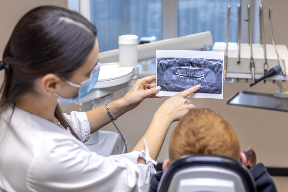
Once your images are ready, Dr. Foleck walks you through what they show on screen. You’ll see exactly where decay, bone changes, or other concerns are located, rather than just hearing about them secondhand. Questions are welcome throughout this part of the visit.

Planning Your Next Steps

Based on what the X-rays reveal, Dr. Foleck outlines what happens from here, whether that’s scheduling treatment, monitoring an area, or simply confirming everything looks healthy. You’ll leave with a clear timeline and understanding of any follow-up imaging needed. Nothing is left vague or open-ended.

Dr. Adam Foleck, DMD, dental implant dentist serving Virginia Beach, Norfolk, and Hampton, VA

Meet the Doctor

Dr. Adam Foleck, DMD, leads the team at The Foleck Center with more than 25 years of clinical experience. He earned his undergraduate degree from the University of North Carolina at Chapel Hill and graduated from Temple University School of Dentistry in 1997, later completing advanced training in general dentistry, implant placement, and bone and gum grafting at the University of Mississippi Medical Center.

He holds Diplomate status in the American Board of Oral Implantology and Honored Fellow status in the American Academy of Implant Dentistry, credentials held by only a small number of dentists nationwide. Dr. Foleck has authored a chapter in the leading textbook Modern Implant Dentistry and lectures internationally on implant surgery and esthetic restoration. He’s supported by a team of skilled doctors, hygienists, and staff who share his commitment to thorough, patient-first care, so every visit to The Foleck Center is backed by decades of combined training and experience.

Learn About Your Doctor!

What Skipping Routine Exams Could Cost You

Choosing not to image or treat a problem right away doesn’t make it disappear, it usually just changes how and when you end up addressing it. Here’s what that trade-off tends to look like in practice.

Financial Implications

Skipping imaging and treating dental problems only when they become painful tends to cost more, not less, over time. A routine dental visit typically runs $90 to $200, while an emergency room visit for the same underlying problem averages around $749 and can climb into the thousands for severe infections requiring hospitalization.

According to the American Dental Association, emergency department visits for dental pain cost roughly three times as much as a routine dental office visit, and preventable dental conditions account for an estimated $1.7 billion in emergency care spending nationally each year.

Health Implications

An untreated cavity or infection doesn’t resolve on its own. Left in place, decay can progress toward the nerve, leading to pain, abscess, and potential tooth loss, while gum disease can quietly damage the bone supporting your teeth long before you notice any symptoms.

One in five working-age adults have at least one untreated cavity, and when conditions like this go unaddressed, the eventual result is often tooth loss rather than resolution.

Safety Implications

An infection that starts in a single tooth doesn’t always stay contained. In rare but serious cases, an untreated dental infection can spread into the jaw, neck, or bloodstream, turning a preventable problem into a medical emergency.

The American Dental Association notes that the most severe untreated dental infections have led to hospitalizations costing hundreds of thousands of dollars, with some cases ending in preventable deaths, underscoring why early detection through routine imaging and exams matters.

FAQs

How much does a dental X-ray cost?

The cost of a dental X-ray depends on the type of image needed and how many views are required for your care. Bitewing and periapical X-rays are typically less expensive than a full panoramic scan, which captures your entire mouth in one image. Many dental insurance plans cover routine X-rays as part of preventive care, so it’s worth checking your benefits or asking our team for a cost estimate before your visit. Your dentist will always explain the recommended imaging and associated cost ahead of time so there are no surprises.

How often should you get dental X-rays?

Most patients need dental X-rays every one to two years, though this can vary based on your oral health history and risk factors. Patients with a history of cavities, gum disease, or ongoing dental work may need more frequent imaging to monitor changes closely. Your dentist will recommend a schedule that fits your specific needs during your regular check-ups.

Are dental X-rays safe?

Yes, dental X-rays are safe, especially with the digital imaging technology used at The Foleck Center, which requires significantly less radiation than traditional film. Every patient is given a lead apron and thyroid collar to further reduce exposure during each scan. The amount of radiation involved is comparable to what you’d naturally encounter in your daily environment over a short period.

Can you get a dental X-ray when pregnant?

Dental X-rays are generally avoided during pregnancy unless there’s an urgent need, such as diagnosing an infection or severe pain that requires immediate attention. If imaging is necessary, extra shielding is used to protect the abdomen and minimize any exposure to the developing baby. Always let your dentist know if you’re pregnant, or think you might be, so the right precautions can be taken before any imaging is scheduled.

Can a dental X-ray show infection?

Yes, dental X-rays can reveal signs of infection, including abscesses, bone loss around a tooth root, and other changes that aren’t visible during a clinical exam. These images help your dentist confirm a diagnosis and determine the right course of treatment quickly, whether that means a root canal, antibiotics, or another approach. Catching an infection early through imaging can prevent it from spreading and causing more serious complications.

Stop Wondering What’s Going On Beneath the Surface

Dr. Adam Foleck and the team at The Foleck Center can get you in for an X-ray and exam this week, often on the same day you call. Dial 757.799.1859 to book directly, or check out real patient stories and behind-the-scenes updates on Facebook, Instagram, TikTok, and YouTube before your visit. Either way, don’t let a hidden problem wait longer than it has to.

Request Appointment